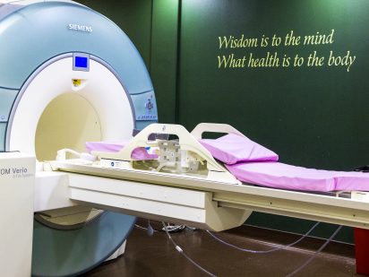
Trinity Health has upgraded its breast MRI program to provide better service to our patients and community by using 3T technology. Now, with the new 3T breast MRI coil, breast cancer that is difficult to see on dense breast tissue can be much better evaluated by our radiologists.
Magnetic resonance imaging, or MRI, uses a magnet and radio waves to produce images of the breasts, to look for tumors and determine whether they’re noncancerous or cancerous, as well as to determine the size and location of a lesion. Trinity Health’s updated technology uses a 3-tesla magnetic field, tesla being a
unit of measurement that measures the strength of a magnetic field. (Trinity Health was the first hospital in the state of North Dakota to invest in a 3T MRI.)
MRI is used in addition to mammography, a screening
process used to check for
breast cancer or other
abnormalities in breast tissue. When a patient gets a mammogram, there may be a suspicious lesion that isn’t examined well enough, despite 2D or 3D mammograms and ultrasound guidance, explained Jim Coffin, RT(R), CT, ARRT, Director of Imaging Services at Trinity Health.
“Occasionally, our radiologists are not able to completely rule out a lesion on 2D or even 3D tomosynthesis mammography,” he said. “As great as 3D mammography is, there are still times when a radiologist needs better information to be confident in their diagnosis and interpretation.”
Most often, a good screening mammogram with 3D is all that is required, however, if the radiologist still has concern, they may order an ultrasound study of a suspicious looking anomaly. Again, most often that will allow the radiologist to be most confident. But for that percentage of patients that have very dense breast
tissue, the radiologist can now suggest breast MRI with 3T strength.
A breast MRI is very much like a standard MRI: a patient lies on the MRI couch and enters the gantry. In this case, the patient would be on their stomach and depending on if they are able, their arms would be over the head. The patient is positioned so the breasts are surrounded by the breast coil. The study lasts about 15 to 20 minutes, during which time contrast and non-contrast images are taken. These images allow the radiologist to search for any “architectural distortion” or a “mass.” Breast MRI is
the most sensitive way of detecting cancers in patients with very dense breast tissue; this is why Trinity Health continues to invest in newer technology.
Additionally, through the 3T technology, needle biopsies can be performed with MRI guidance, Coffin said. The biopsy material is reviewed by a pathologist, who would report the results back to the referring radiologist and ordering provider.
Technically speaking:
• Higher magnetic field strength (3T) improves quality of breast MRI imaging
• Higher resolution imaging assists in characterizing breast lesions
• Breast MRI detects small cancers earlier with greater detail
• MRI imaging provides the ability to use contrast enhancement to highlight tumor activity
• MRI allows excellent discrimination between malignant vs non-malignant masses (cancer or non-cancer)
Practically speaking:
• 3T breast imaging allows the radiologist to better examine dense breast tissue
This new 3T program was a request by Brian Johnson, DO, Trinity Health’s breast fellow trained radiologist. Dr. Johnson earned a Master of Science in Physician Assistant Studies from Philadelphia College of Osteopathic Medicine. He received his Doctor of Osteopathic Medicine from Lake Erie College of Osteopathic Medicine in Pennsylvania. Following a transitional internship at Sanford Health – University of North Dakota School of Medicine, Fargo, he completed a four-year residency in Diagnostic Radiology at Strong Memorial Hospital – University of Rochester School of Medicine, Rochester, NY. He went on to complete his breast imaging fellowship at New York University Langone Health Center in New York, NY. Dr. Johnson is a member of the Radiological Society of North America, American College of Radiology, American Roentgen Ray Society, and Society of Breast Imaging.

